
Physics and physical properties to be considered when utilizing therapeutic laser energy for bio modulation of cellular and tissue function.
Written by Dr. Peter H. Eeg BSc, DVM, CVLF, FASLMS
Poolesville Veterinary Clinic, Poolesville, Maryland

Physics and physical properties to be considered when utilizing therapeutic laser energy for bio modulation of cellular and tissue function.
Written by Dr. Peter H. Eeg BSc, DVM, CVLF, FASLMS
Poolesville Veterinary Clinic, Poolesville, Maryland
Written by Dr. Peter H. Eeg, BSc, DVM, CVLF, FASLMS
Poolsville Veterinary Clinic
Written by Dr. Peter H. Eeg, BSc, DVM, CVLF, FASLMS
Poolsville Veterinary Clinic
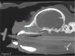
Small Animal Case Review by Dr. Bob O'Brien, DVM, DACVR
How is Artemis™ different from other laser therapy devices currently available for veterinary use?
There is only minimally superficial similarities between Artemis™ and previous therapeutic lasers. Previous lasers were diode lasers and Low Level Laser Therapy (LLLT). Artemis™ is by far the most advanced High Intensity Laser Therapy device in the world.
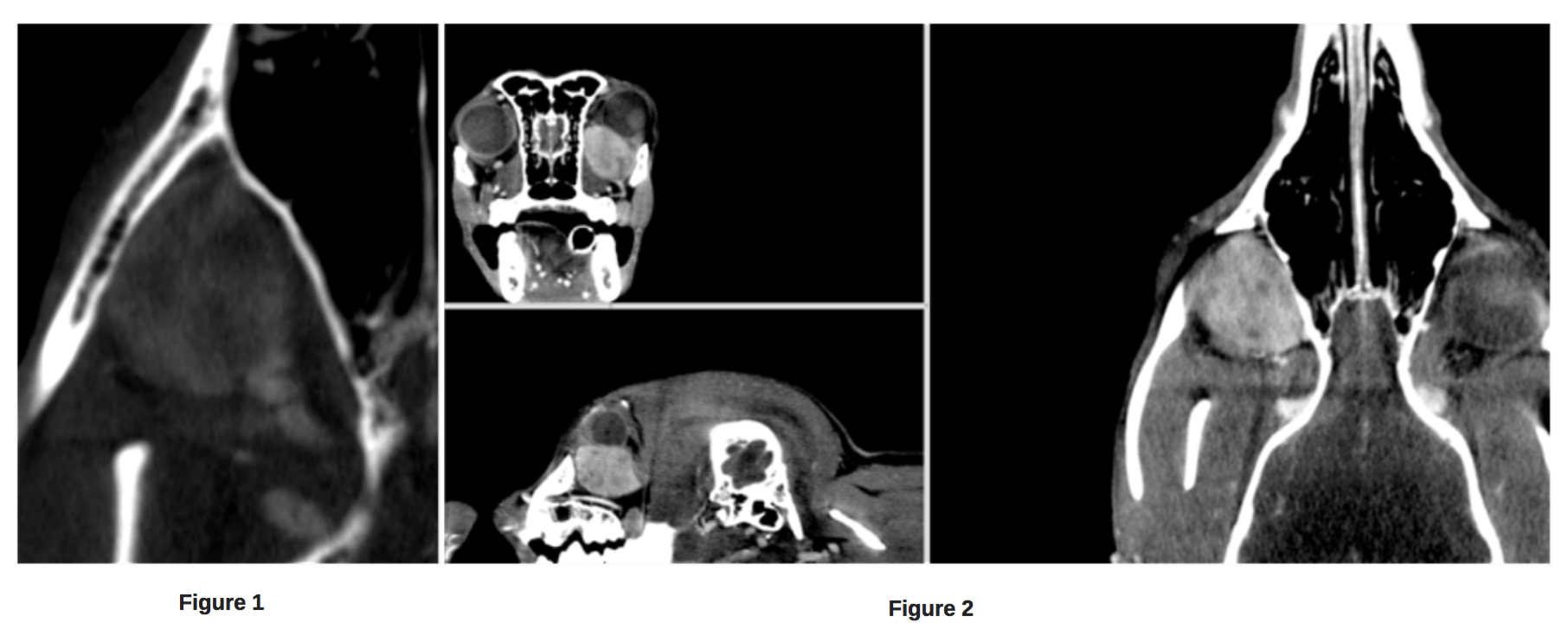
Small Animal Case Review by Dr. Bob O'Brien, DVM, DACVR
9-year-old female spayed Border Collie that presented to a surgical specialty practice with a 6 month history of left sided exophthalmos.
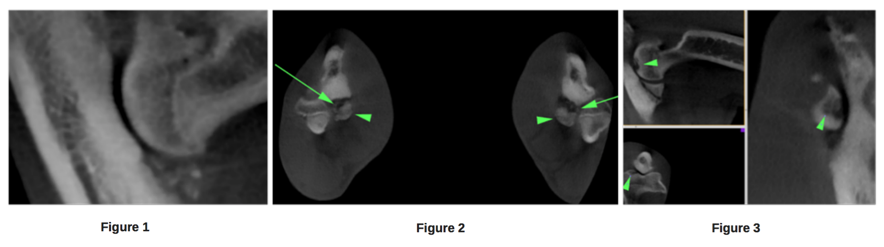
Small Animal Case Review by Dr. Bob O'Brien, DVM, DACVR
9-month-old male neutered Labrador retriever dog that presented with acute onset 3 days previously, of right forelimb lameness. On physical examination, bot elbows were painful on flexion and extension.
Small Animal Case Review by Dr. Bob O'Brien, DVM, DACVR
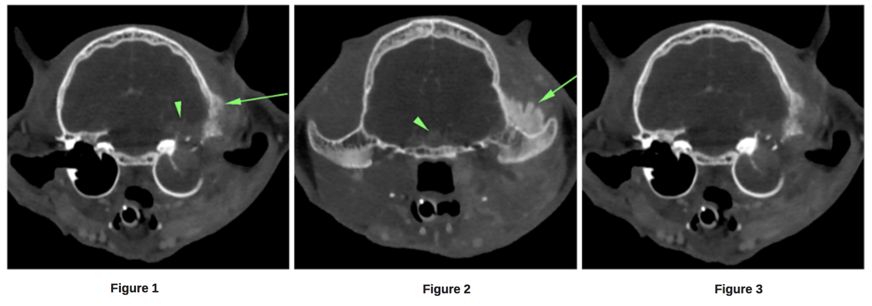
4 year old male neutered domestic short haired cat that presented with a history of chronic ear scratching. Recently the patient developed a head tilt. On physical examination the patient had soft tissue swelling to the left base of the skull, head tilt, and nystagmus. The left ear canal was occluded at the junction of the horizontal and vertical ear canal regions.
Small Animal Case Review by Dr. Bob O'Brien, DVM, DACVR
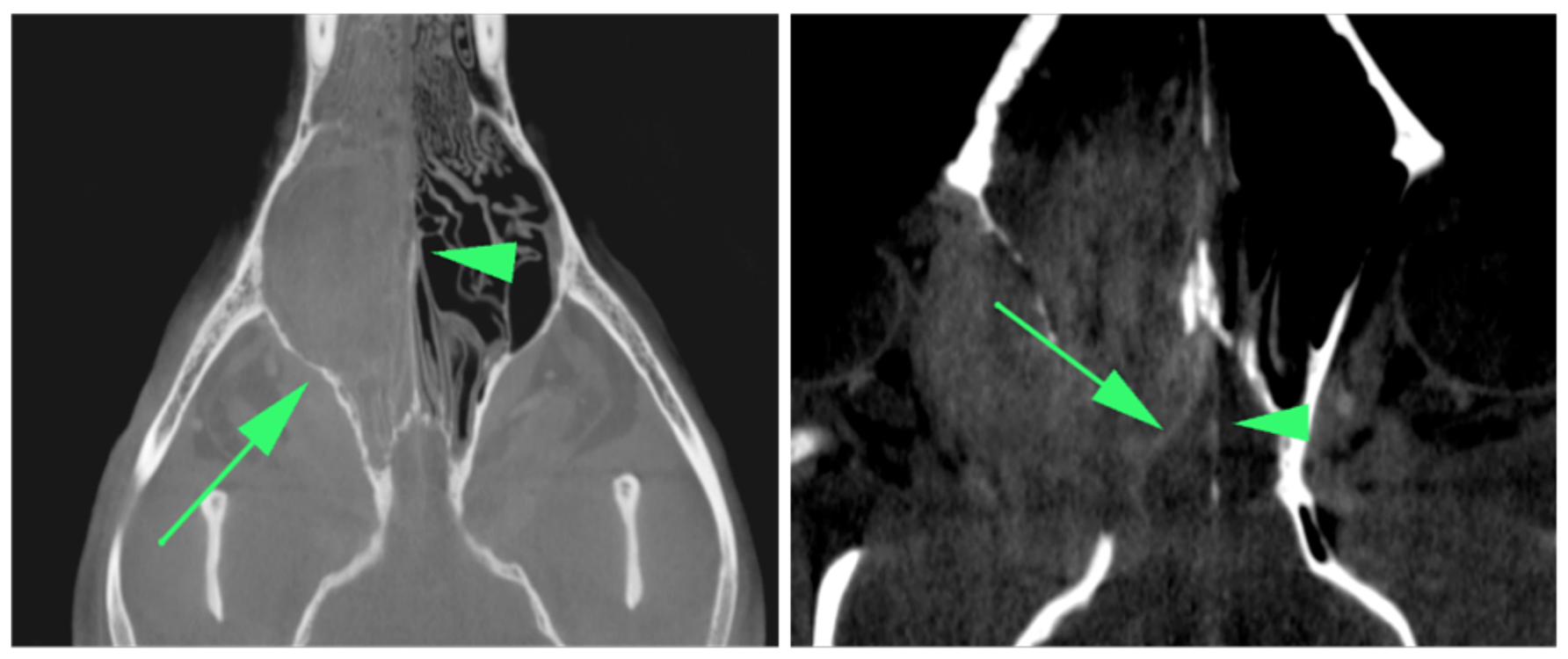
10-year-old male neutered welsh terrier dog that presented with a 2-month duration history of right sided mucopurulent nasal discharge that recently became bloody.
Small Animal Case Review by Dr. Bob O'Brien, DVM, DACVR
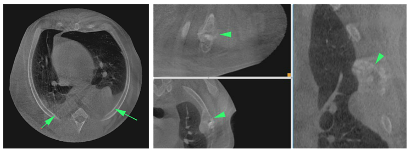
6-year-old male neutered large mixed breed dog that presented with a history of acute lethargy and labored breathing. Thoracocentesis revealed hemothorax. Image protocol: The patient was imaged in dorsal recumbency. A survey and post-i.v. contrast CT was performed of the chest. The contrast dose was 1 ml/lb BW of Iohexol (300mg/ml iodine) contrast media; given as a bolus prior to image initiation.