
Small Animal Case Review by Dr. Bob O'Brien, DVM, DACVR
Patient
10-year-old male, neutered Cavalier King Spaniel.
History
Choking, gasping, coughing, difficulty eating, bark change for several days.
Image Protocol
A survey and post- i.v. contrast CT study was performed of the nasal cavity and skull.
Findings
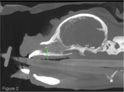
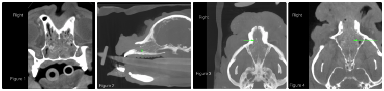
Increased soft tissue material was seen in both nasal cavities (Figure 1). Normal turbinates were noted within this affected region without evidence of lysis or mass effect. A small (6 mm long by 1 mm wide and tall) mildly boney lesion was seen in the mid-rostral right nasal cavity (Figures 2 & 3). No mass lesions was seen to be associated with this linear-shaped lesion. Mild undulation of the septum was noted as a normal= breed variant (Figure 4). The right tympanic bulla was filled with soft tissue material and there was no evidence of either lysis or proliferation in the bulla wall (Figure 5). The left tympanic bulla was normal. Mild multifocal mineralization was seen in the horizontal and vertical ear canals bilaterally.
Imaging Diagnosis
Nasal cavity: possible foreign material (or atypical dystrophic mineralization) Right tympanic bulla: otitis media (mild, acute).
Final Diagnosis
Blades of grass were removed from the right nasal cavity on rhinoscopic evaluation. The otitis resolved on empirical antibiotics. The patient is now clinically normal.
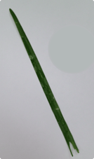
This case is an excellent example of nonaggressive changes seen very well on the Vimago™ system due the exceptionally high spatial resolution. The excellent contrast resolution allowed identification and characterization of the grass material.
Discussion
CT is very sensitive to changes within the nasal cavities. The assessment of aggressive (versus nonaggressive) changes in the nasal cavity is based on presence of turbinate destruction, boney septum deviation or lysis, mass effect, and lysis of surrounding nasal vault. Staging the mass often hinges on presence of brain involvement. Non aggressive lesions include immune mediated, infectious (especially fungal), dental and foreign body causes. This case is an excellent example of nonaggressive changes seen very well on the Vimago™ system due the exceptionally high spatial resolution. The excellent contrast resolution allowed identification and characterization of the grass material.
Special Thank you to Dr. Lyman and his team @ Animal Emergency and Referral Clinicnimal Emergency and Referral Clinic in Ft. Pierce, FL. Learn more abour AERC here.
 What sets Vimago™ apart from other CT devices? Watch our short video.