
Small Animal Case Review by Dr. Bob O'Brien, DVM, DACVR
PATIENT
6-year-old male neutered large mixed breed dog that presented with a history of acute lethargy and labored breathing. Thoracocentesis revealed hemothorax. Image protocol: The patient was imaged in dorsal recumbency. A survey and post-i.v. contrast CT was performed of the chest. The contrast dose was 1 ml/lb BW of Iohexol (300mg/ml iodine) contrast media; given as a bolus prior to image initiation.
FINDINGS
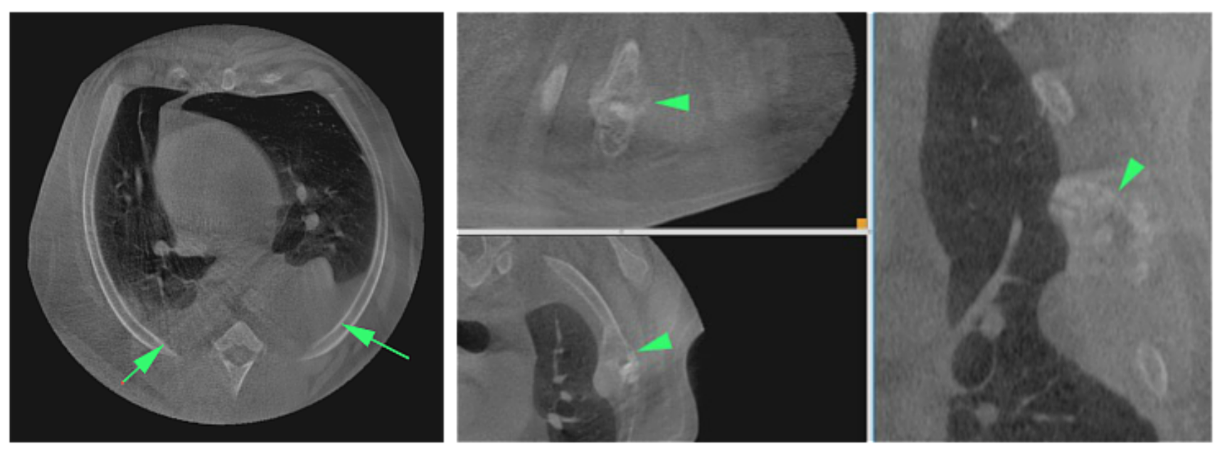
A lesion was seen in the distal aspect of the left 4th rib. The lesion had moth eaten lysis, irregular, speculated periosteal proliferation and a pathological fracture with minimal displacement. An extrapleural sign was seen with the adjacent lung. Soft tissue attenuation material is seen bilaterally in the dependent (dorsal) region bilaterally (left worse than right) .
IMAGING DIAGNOSIS
Primary rib neoplasia (OSA, CSA, atypical myeloma, metastatic lesion or osteomyelitis) with pathological fracture and secondary hemothorax.
"The Vimago™ HD CT is and excellent choice for imaging all intrathoracic diseases."
DISCUSSION POINT
This patient demonstrated the ability of the Vimago™ Veterinary CT Scanner to image larger patients. The spatial resolution of the rib and pleural space lesions were excellent. The ability to scan the remaining lung for metastatic lesions was excellent. We prefer to image the chest of dogs with the patient in dorsal recumbency since the inevitable mild movements are less evident than with the patient in ventral (sternal) recumbency. Even though 3 separate image acquisitions were required to cover the entire chest length, there is no evidence of the splicing that occurs to put the 3 sets of images together into one file. The Vimago™ HD CT is an excellent choice for imaging all intrathoracic diseases.

Confused about the difference between standard CT and HD CT? Watch our short video below.