
Small Animal Case Review by Dr. Bob O'Brien, DVM, DACVR
PATIENT
9-month-old male neutered Labrador retriever dog that presented with acute onset 3 days previously, of right forelimb lameness. On physical examination, bot elbows were painful on flexion and extension.
IMAGE PROTOCOL
Survey CT was performed of both elbows.
FINDINGS
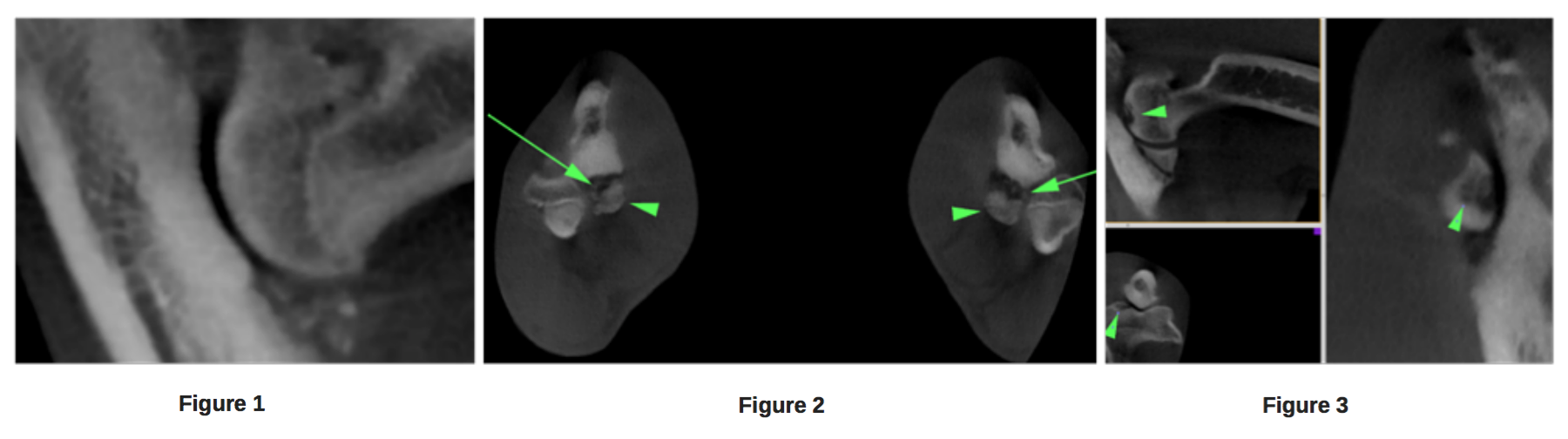
Both elbows had similar findings. The was a large separate fragment cranial to the cranial aspect of the medial coronoid process of the ulna (FIG 1). Additionally, many small fragments (0.2 – 0.8 mm wide) were seen at the base of the larger fragments (FIG 2). The subcondral bone of the semilunar notch of the ulna was sclerotic. Osteophytes were seen on the medial and lateral aspects of the distal humerus, proximal aspect of the anconeal process and cranial border of the radial head. Irregular shaped subcondral lysis was seen surrounded by sclerosis in the medial condyle articular surface (FIG 3).he entire right nasal cavity and frontal sinus was filled with uniform soft tissue opacity, mildly enhancing material. There is indistinct turbinate destruction and deviation of the boney septum to the left. There was lysis on the left side of the nasal vault; maxilla, nasal and adjacent caudal bones. Lysis of the cribriform plate was seen with the mass invading into the brain causing deviation of the contrast enhanced meninges and mild leftward deviation of the contrastenhanced falx cerebri.
IMAGING DIAGNOSIS
Bilateral elbow degenerative joint disease cause by elbow dysplasia: a. fragmented medial coronoid process b. osteochondrosis of the medial condyle.
"The Vimago™ HD CT is the perfect imaging system for charavterization of developmental and acquired bone and joint diseases."
DISCUSSION POINT
Elbow imaging is the most common musculoskeletal CT study in young lame canine patients. This is because of the inherent insensitivity of conventional radiography (or ultrasound) for imaging the medial coronoid process of the ulna due to superimposition of adjacent structures. CT provides high detail crosssectional imaging without the disadvantages of superimposed structures. The Vimago™ veterinary CT scanner has excellent spatial resolution, much better than conventional CT, allowing accurate measurement of fragments of 0.2 mm in this case. Elbow studies are very easy to perform with the Vimago™ HD CT system. The forelimbs are extended to approximately the same level and a single image collection set is made. No contrast is necessary for these types of studies. The subsequent data set can easily be manipulated by the reviewer into any image plane necessary to identify all the pertinent structures of the bones of the elbows. The Vimago™ HD CT is the perfect imaging system for characterization of developmental and acquired bone and joint diseases.

Confused about the difference between standard CT and HD CT? Watch our short video below.