
Small Animal Case Review by Dr. Bob O'Brien, DVM, DACVR
PATIENT
12-year-old male neutered mix breed dog that presented with a 3month duration history of hematuria. The prostate was enlarged on rectal examination.
IMAGE PROTOCOL
A survey and posti.v. contrast CT was performed of the abdomen. The contrast dose was 1ml ml/lb BW of Iohexol (300mg/ml iodine) contrast media given as a bolus immediately prior to image initiation.
FINDINGS
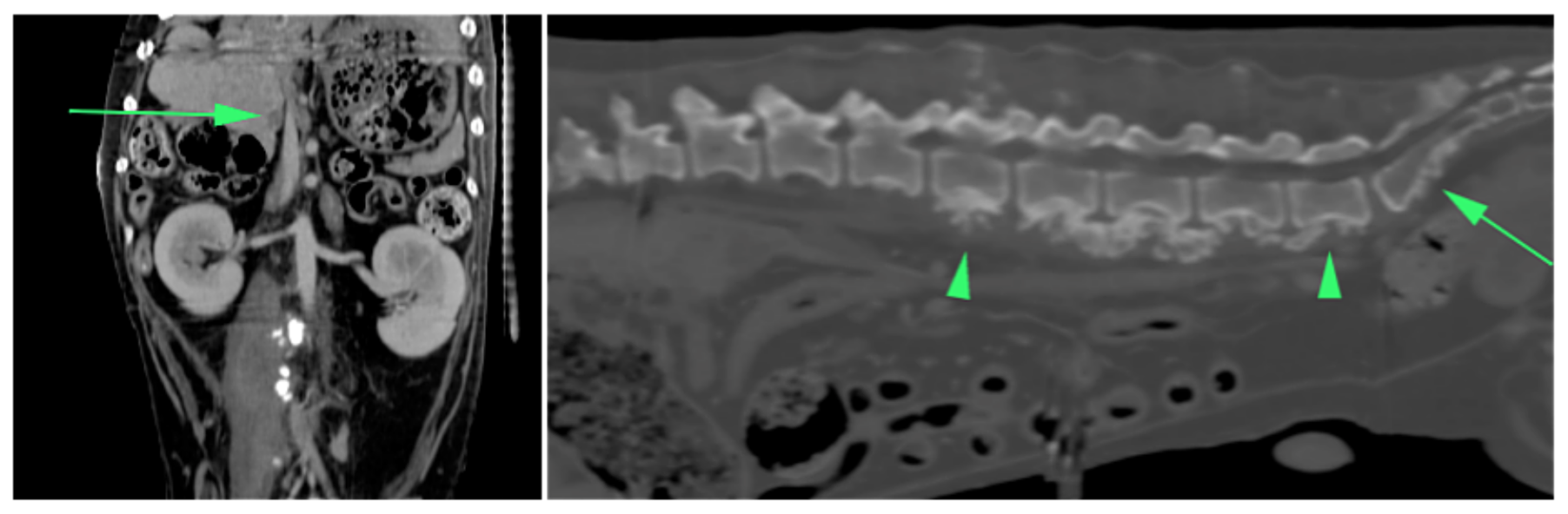
The prostate was moderately enlarged with patchy regions of poor contrast enhancement centrally (FIG 1). The overall shape and contour was normal. The medial iliac lymph nodes were normal shape and size. On survey CT the liver had an undulating outer contour, but no nodules were seen. On postcontrast CT images, poorly enhancing nodules (48mm wide) were seen in all liver lobes. The lumbar spine has very active appearing, speculated periosteal proliferation on the ventral aspect of the vertebral bodies from L2L7 (FIG 2). This proliferation seemed to originate from the central portion of the bodies. No lysis was seen. A similar appearance was seen on the ventral aspect of the sacral vertebrae and both ilial wings.
IMAGING DIAGNOSIS
Liver: metastastic neoplasia Spine: neoplastic reactive periosteal changes (or atypical metastasis) Prostate: carcinoma (or abscessation).
DISCUSSION POINT
Staging complicated intraabdominal lesions can be very challenging with conventional radiography and ultrasound. Each modality has their advantages, but neither, even when combined, provides the ability to provide overall high spatial resolution of all bones and intraabdominal organs, provide contrast enhancement information (vital for assessment of nodular lesion in the liver) and high detail cross sectional bone detail. The Vimago™ veterinary CT scanner provided all three of these vital attributes. Because this was a large dog, the acquisition was made of three separate, sequential overlapping image acquisitions automatically spliced into one data set. No other system can provide this function with the ability to image at extremely high spatial resolutions (< 200 uM). The contrast resolution of the postcontrast images provided the basis for identifying the liver nodules (not seen on the ultrasound scan) and characterization as probably malignant.

Confused about the difference between standard CT and HD CT? Watch our short video below.